Clinical-grade AI tools for every stage of brain health
From early detection to longitudinal monitoring. Quantitative neuroimaging that fits into existing clinical workflows.
Alzheimer's Early Detection
Cedawn, our Alzheimer's platform, is live. It performs quantitative volumetric analysis of clinically relevant brain regions associated with Alzheimer's disease progression. Upload a standard brain MRI and receive a clinician-ready report showing region-by-region measurements, normative comparisons, and staging of structural changes.
- 10 clinically relevant brain regions analyzed per scan
- Normative comparison against age-matched controls
- Longitudinal tracking for disease progression
- Standard medical imaging format support
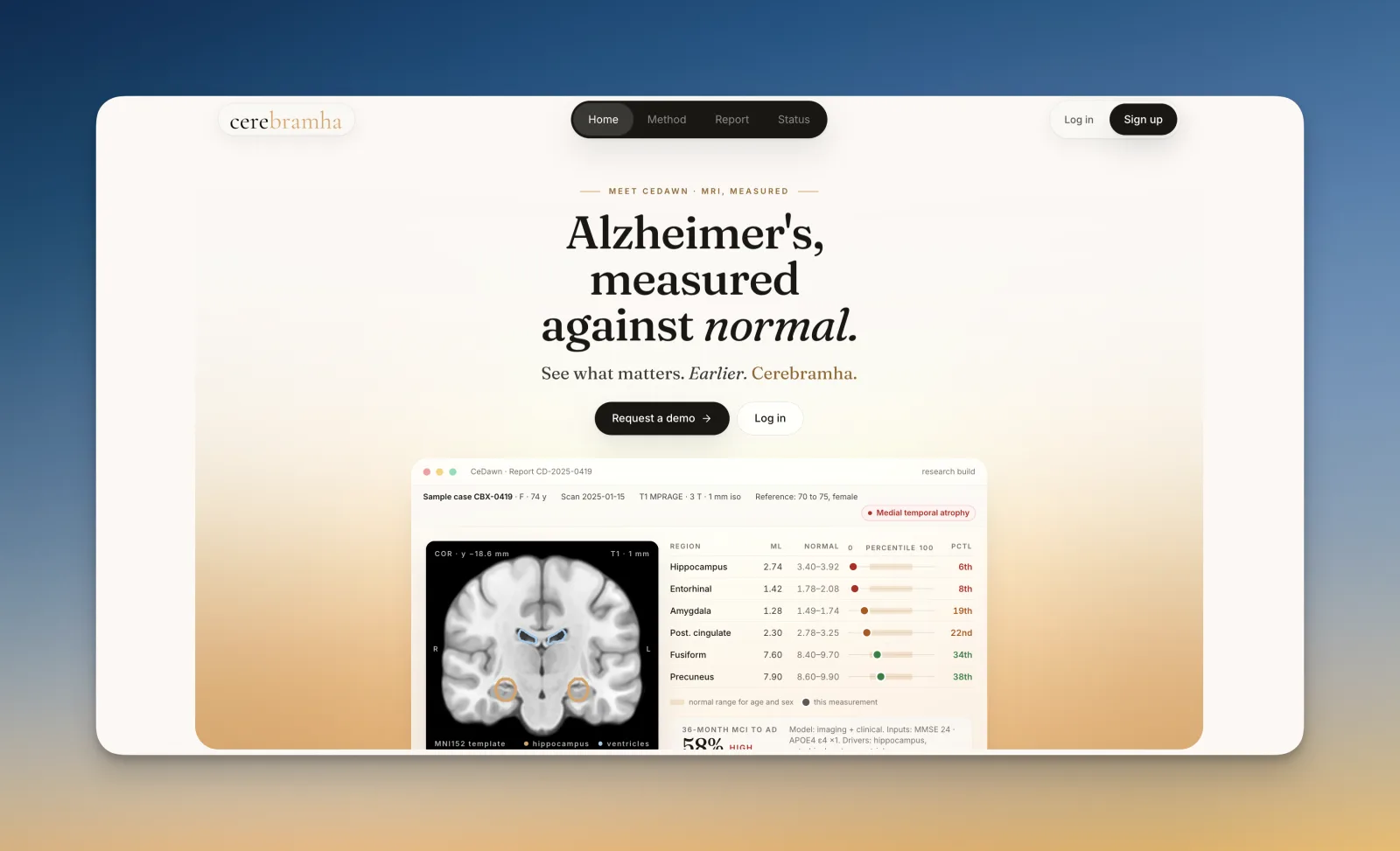
Three steps from scan to clinical insight
Upload Scan
Upload a brain MRI scan from any clinical scanner. No special hardware or preparation required.
AI Analysis
Our AI engine automatically prepares and analyzes the scan, segmenting clinically relevant brain regions with precision.
View Results
Volumetric measurements compared against normative data matched by age and sex. Downloadable clinical report.
The ten regions we measure
Each is segmented and measured against normative data matched by age and sex.
Hippocampus
Memory formation. Among the first structures affected in Alzheimer's disease.
Entorhinal Cortex
Gateway to hippocampus. Critical for spatial memory and early-stage degeneration.
Amygdala
Emotional processing. Atrophy correlates with behavioral and psychological symptoms.
Inferior Temporal Gyrus
Visual object recognition. Shows early cortical thinning in Alzheimer's.
Frontal Lobe
Executive function and planning. Affected in moderate-to-late stages.
Parietal Lobe
Spatial awareness and sensory integration. Atrophy impacts daily functioning.
Posterior Cingulate
Default mode network hub. Early metabolic changes visible on PET.
Precuneus
Self-referential processing. Early amyloid deposition site.
Thalamus
Sensory relay center. Volume loss indicates broader network disruption.
Cerebellum
Motor coordination. Used as a reference region for normalization.
Region-Based
Alzheimer’s Risk Analysis.
Four of the ten measured regions, and what their structural change indicates.
Hippocampus
Most Important Early Marker
The hippocampus is critical for memory formation and spatial navigation. Early hippocampal atrophy is one of the strongest structural predictors of Alzheimer’s disease.
Clinical note
Hippocampal shrinkage often appears before severe cognitive symptoms, making it central to early-stage detection models.
Structural signals analyzed
Volume reduction patterns
Asymmetry between hemispheres
Early microstructural signal deviations
Inferior Temporal Gyrus
Progression Indicator
The inferior temporal gyrus supports visual object recognition and semantic memory. Cortical thinning here is closely associated with progression from Mild Cognitive Impairment to Alzheimer’s.
Clinical note
Inferior temporal thinning correlates with measurable decline in naming and object-recognition tasks.
Structural signals analyzed
Cortical thinning
Structural integrity mapping
Signal density variation
Parietal Lobe
Cognitive Integration
The parietal lobe contributes to attention, orientation, and integration of sensory information. Advanced Alzheimer’s progression often involves parietal structural disruption.
Clinical note
Changes in this region are associated with spatial disorientation and impaired decision-making.
Structural signals analyzed
Pattern disorganization
Connectivity disruption indicators
Regional signal attenuation
Amygdala
Supportive Marker
The amygdala is involved in emotional regulation and memory processing. Structural changes here may support early differential diagnosis.
Clinical note
Amygdala involvement can provide additional context in distinguishing Alzheimer's from other neurodegenerative conditions.
Structural signals analyzed
Subtle volume deviation
Connectivity anomalies
Multi-modal feature coupling
Every region is measured volumetrically and compared against normative data matched by age and sex, so structural change is read against a baseline rather than in isolation.
What we are building next
Brain Tumour Segmentation
Automated detection, precise volumetric segmentation, and classification of brain tumors for targeted surgical planning and treatment monitoring. In development. Not cleared for diagnostic use.
Built for clinicians and researchers alike
Built for the radiology workflow
- PACS integration (Orthanc-compatible)
- Standard PDF report format
- Sub-5-minute report turnaround
- No additional hardware required
- Normative comparison by age and sex
- Designed for radiologist workflow
Be part of building this
The research API is not open yet, because we are still building it. If you work in neuroimaging, computational neurology, or clinical AI and want to shape what this becomes, we would genuinely love to have you involved.
What we are planning
- API access for batch processing (planned, not open yet)
- Standard medical imaging format support
- Raw analysis outputs available
- Longitudinal analysis built in
- Programmable report generation
We'd love to have you
If you work in neuroimaging, clinical AI, or computational neurology, reach out. Early collaborators help define the direction. We are grateful for every person who believes in this early.
- Shape the research API from day one
- Early access before public launch
- Direct line to the founding team
- Co-author research if applicable